GET /conditions/77/?format=api
HTTP 200 OK
Allow: GET, PUT, PATCH, DELETE, HEAD, OPTIONS
Content-Type: application/json
Vary: Accept
{
"id": 77,
"url": "https://cclm.media-doc.io/conditions/77/?format=api",
"name": "E. Conical beam",
"category": "Biomicroscope",
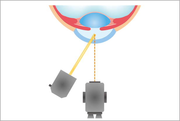
"detail": "<h2>Overview</h2>\n\n<ul>\n\t<li>A conical beam is useful for assessing the anterior chamber and anterior vitreous</li>\n\t<li>It is used to look for the presence of either cells (flare) or pigment in the aqueous or vitreous</li>\n</ul>\n\n<p> </p>\n\n<h2>Set Up</h2>\n\n<ul>\n\t<li>Room lights should be turned off and time given for the practitioner to dark adapt</li>\n\t<li>The light beam is adjusted to a narrow width</li>\n\t<li>The height of the beam is reduced until it forms a small circular shape</li>\n\t<li>High magnification is used</li>\n\t<li>The biomicroscope is focussed on the anterior chamber or anterior vitreous</li>\n\t<li>Detection of cells or pigment is enhanced by asking the patient to move their eyes and then watching for any movement as they look straight ahead again </li>\n</ul>\n\n<p> </p>\n\n<h2>Useful when assessing</h2>\n\n<ul>\n\t<li>Looking for signs of active inflammation - flare (Tyndall effect)</li>\n\t<li>Looking for signs of retinal detatchment - pigment (Schaeffer's sign)</li>\n</ul>\n\n<p> </p>",
"images": [
{
"name": "Conical beam",
"file": "https://cclm-static.s3.amazonaws.com/images/CORE_Conical.png"
}
],
"type": 1,
"videos": [
{
"name": "Conical Beam",
"videoSrc": "267427065"
}
],
"wear": false,
"ceaseWear": false,
"changeLens": false,
"changeSolutions": false,
"changeCare": false,
"counseling": false,
"dd": false,
"dw": false,
"gp": false,
"replace": false,
"review": false,
"rewettingDrops": false,
"rx": false,
"SiHy": false,
"toric": false,
"tags": [
{
"id": 7,
"url": "https://cclm.media-doc.io/tags/7/?format=api",
"name": "conditions"
}
]
}